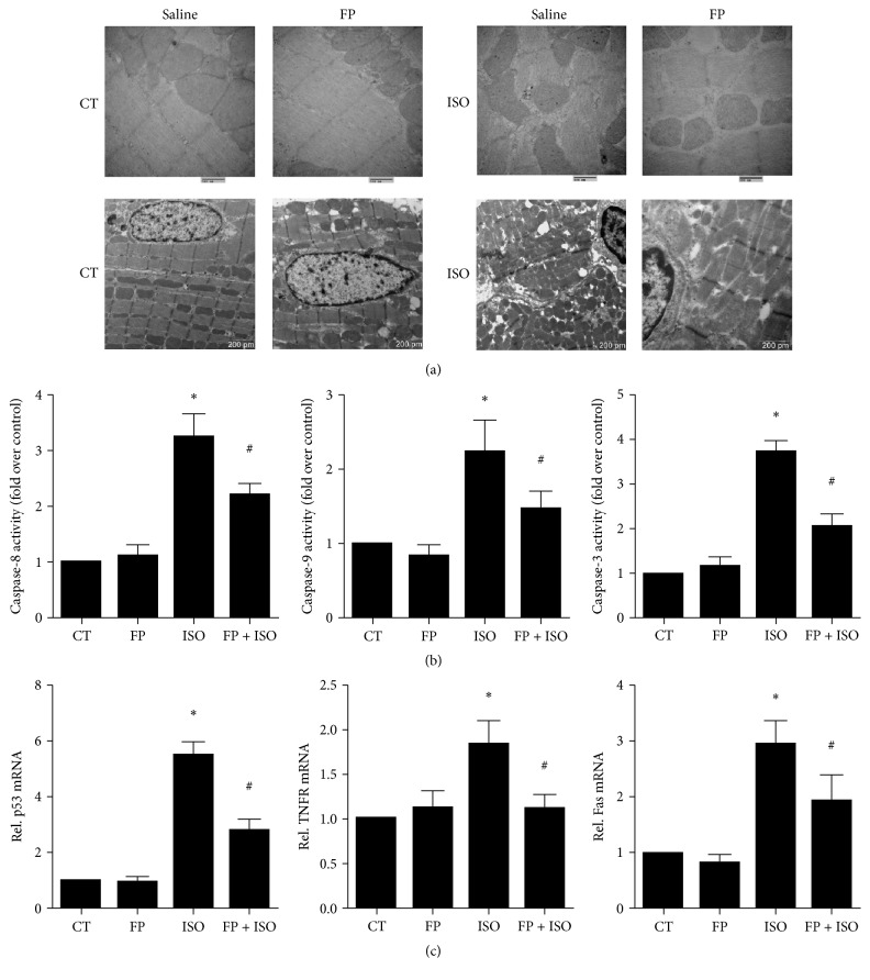
Figure 5.
Effects of isoproterenol and FP on apoptotic damage and apoptosis-related gene expression in heart tissues. (a) Transmission electron microscopy of heart tissues. (b) The activities of caspase-8, caspase-9, and caspase-3 were measured using a fluorometric assay and expressed as the fold change over the control. (c) mRNA levels of p53, TNFR1, and Fas were determined by real-time RT-PCR. The levels of mRNA were normalized to GAPDH. Relative mRNA levels are shown using arbitrary units, and the value of the control group (CT) is defined as 1. The results are expressed as the means ± SE; n = 10 mice per group (∗ p < 0.05 compared to the control group; # p < 0.05 compared to the isoproterenol group).

