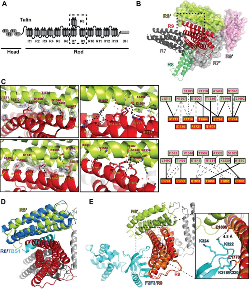
Figure 1. Structure of talin R7R8R9.

(A) Schematic diagram of talin. The head region possesses the F0, F1, F2, and F3 domains (ovals). The rod region contains 13 helical bundle domains (R1–R13) and a dimerization helix (DH). The R7R8R9 fragment is indicated by the dashed box. (B) Cartoon diagram of wild-type talin triple domains R7R8R9. The R7, R8, and R9 domains are colored in dark gray, green, and red, respectively. A symmetrically related R7R8R9 molecule is shown in surface representation (R7’ in light gray, R8’ in lime, and R9’ in pink). The interface formed by the R9 and R8’ domains is indicated by the dashed box. (C) Close-up views of the dashed box from (B) in both front view (upper panels) and back view (lower panels). Hydrogen bonds are indicated by dotted lines and van der Waals contacts are represented by surfaces. Interacting residues are labeled (R8’ in red-black text and R9 in yellow-black text). Schematic maps of the interactions are shown on the right with van der Waals contacts in solid line, direct hydrogen bonds in dotted lines, and water-mediated hydrogen bonds in gray dashed lines. (D) The crystal structure of R8:TBS1 (PDB ID: 4W8P. R8 in dark blue, TBS1 in cyan) is superimposed on the symmetrically related R8’ domain (lime) of the R7R8R9 structure. The TBS1 fragment sterically clashes with the R9 domain (red). (E) The crystal structure of F2F3:R9 (PDB ID: 4F7G. R9 in salmon, F2F3 in cyan) is superimposed on the R7R8R9 structure. The salt bridge interactions of K318/K320 in the lysine finger of the F3 domain and E1770 in the R9 domain are shown in the inset box. The amino group of Lys322 and the carboxyl group of Glu1805 is 4.8 Å apart, and thus does not form a salt bridge.
