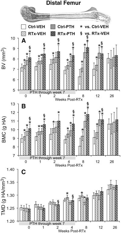
Fig. 4.
Micro-CT analysis of the distal 5-mm femur volume of interest. a The distal femur bone volume (BV) was increased in irradiated groups through week 12, though to a greater magnitude in the RTx-PTH group than the VEH-RTx group. b Treatment effects on bone mineral content (BMC) paralleled BV responses. c Bone tissue mineral density (TMD) was transiently increased in the RTx-VEH group at weeks 4–8. Data are graphed as average ± standard deviation, with statistical significance indicated at t < 0.05 for RTx-VEH vs. Ctrl-VEH or p < 0.05 for all other comparisons

