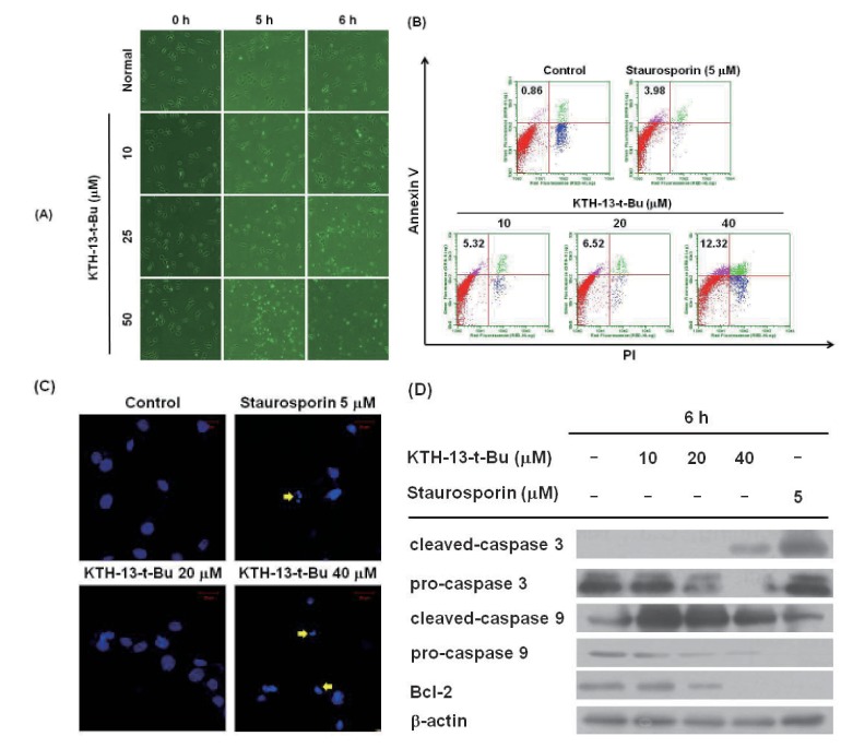
Fig. 3. Effect of KTH-13-t-Bu on inducing apoptosis in C6 glioma cells.
(A) C6 glioma cells (5×10⁵ cells/ml) were incubated with KTH-13-t-Bu for 0, 5, and 6 h. Morphological chan ges were detected at each time point by microscopic analysis. (B) Dose-dependent pro-apoptotic effect of KTH-13-t-Bu was measured by FITC annexin V-PI staining assay. Cells were treated with annexin V, PI, and KTH-13-t-Bu (0 to 40 µM) for 6 h. Stained cells were detected by flow cytometry. (C) Changes the nuclear morphology of C6 glioma cells induced by KTH-13-t-Bu were analyzed by confocal microscopy after DAPI staining. (D) Dose-dependent upregulation of apoptosis-related proteins induced by KTH-13-t-Bu. C6 glioma cells (5×10⁵ cells/ml) were incubated with KTH-13-t-Bu for 6 h. The levels of total and cleaved caspase 3, caspase 9, and Bcl-2 were detected by immunoblotting analysis.

