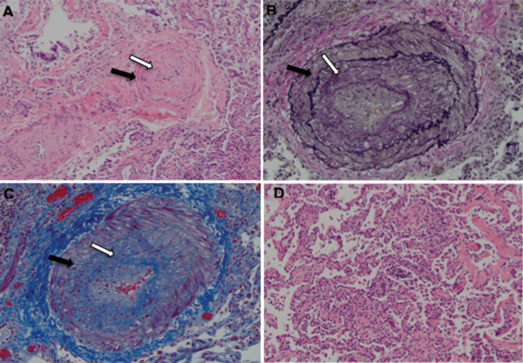
Figure 2.
Lung biopsy demonstrating pulmonary arterial hypertension and patchy nonspecific interstitial pneumonia. A, Hematoxylin-eosin stain (×200) showing a nearly occluded pulmonary artery branch with smooth muscle hypertrophy (black arrow) and intimal proliferation (white arrow). B, Elastin stain (×200) showing widening of the space between the internal and external elastica by smooth muscle hypertrophy (black arrow) as well as intimal proliferation (white arrow). C, Trichrome stain (×200) showing red smooth muscle hypertrophy (black arrow) and blue fibrous tissue in the intima (white arrow). D, Hematoxylin-eosin stain (×200) showing a segment of interstitial inflammation. No interstitial fibrosis was seen, and no venous thrombi or plexiform lesions were seen.

