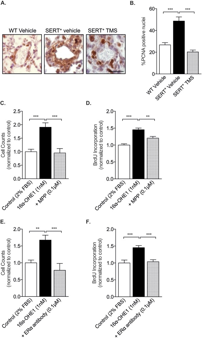
Figure 8.

The cytochrome P450 1B1 (CYP1B1) metabolite 16α-hydroxyestrone (16α-OHE1) induces cell proliferation via estrogen receptor α (ERα). A, Representative images of immunohistochemistry analysis of the DNA replication marker PCNA in wild-type (WT) mice and in SERT+ mice treated with vehicle or the CYP1B1 inhibitor 2,3′,4,5′-tetramethoxystilbene (TMS). Scale bar: 20 μm. B, Percentage PCNA-positive nuclei; n = 3–6, 3 or 4 pulmonary arteries analyzed per lung. Data were analyzed by a 1-way analysis of variance (ANOVA) followed by a Tukey post hoc test. C, D, Female human pulmonary arterial smooth muscle cells (PASMCs) were incubated with 16α-OHE1 for 72 hours in 2% charcoal-stripped serum in the presence or absence of ERα inhibition and assessed for proliferation by cell counts and BrdU incorporation. 16α-OHE1 induced PASMC proliferation in the presence of a selective ERα antagonist MPP dihydrochloride (0.1 μM), as assessed by cell counts, n = 15 replicates per group (C), and BrdU incorporation, n = 11 replicates per group (D). E, F, 16α-OHE1 induced PASMC proliferation in the presence of a specific ERα antibody (0.002 pg/mL) as assessed by cell counts, n = 6–9 replicates per group (E), and BrdU incorporation, n = 16 replicates per group (F). Data were analyzed by a 1-way ANOVA followed by a Tukey post hoc test. **P < 0.01; ***P < 0.001. BrdU: bromodeoxyuridine; FBS: fetal bovine serum; PCNA: proliferating cell nuclear antigen; SERT+: serotonin transporter is overexpressed.
