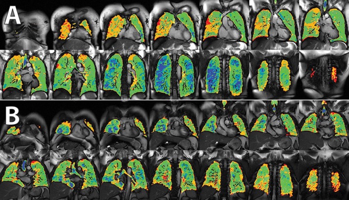
Figure 4.
Differences in ventilation imaging between subjects with pulmonary vascular disease (PVD; A) and idiopathic pulmonary fibrosis (IPF; B). Subjects with PVD appear to have fairly normal ventilation with relatively high lung compliance (blue), while those with IPF appear to lose high-intensity signal (loss of blue signal), consistent with a loss of lung compliance.

