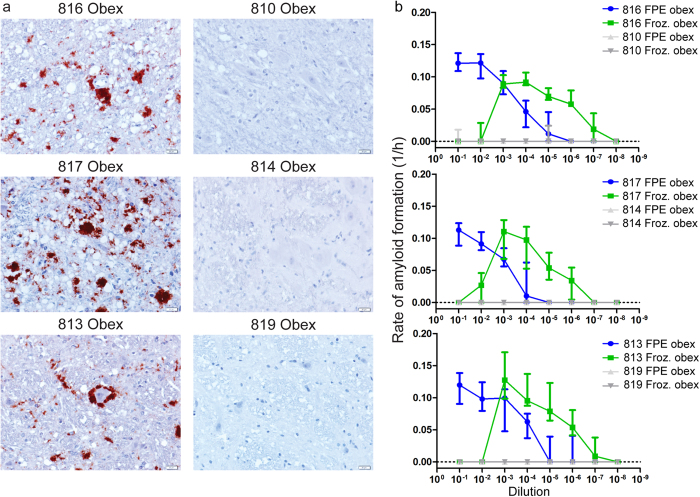
Figure 2. Detection of amyloid seeding activity in FPE obex tissue.
(a) Immunohistochemistry of obex from three terminal white-tailed deer. All display variably sized PrPCWD plaque accumulations in the neuropil. The corresponding negative control animals do not display any PrPCWD immunoreactivity. Images are 400× magnification, measurement bars represent 20 μ m. (b) RT-QuIC rates of amyloid formation of serial dilutions of FPE tissues from IHC positive obex and frozen 10% obex tissue homogenate. FPE samples from IHC positive obex are detectable in RT-QuIC from all three white-tailed deer examined. The RT-QuIC linear range in FPE obex samples extends from 10−1 to 10−6 whereas the linear range in frozen 10% homogenates is from 10−4 to 10−7. Spontaneous amyloid formation from rPrPC negative control samples occurred equally rarely in both FPE and frozen homogenates. Each point represents median and interquartile range (IQR) and is derived of 8 replicates from a minimum of two separate experiments.

