Figure 4.
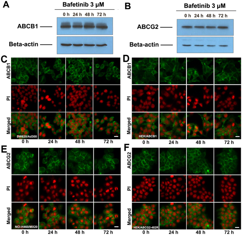
(A)The effect of bafetinib at 3 μM on the expression levels of ABCB1 in SW620/Ad300 cells for 24, 48 and 72 h. (B) The effect of bafetinib at 3 μM on the expression level of ABCG2 in NCI-H460/MX20 cells for 24, 48 and 72 h. Equal amounts of total cell lysate were used for each sample. The effect of bafetinib at 3 μM on the subcellular localization of ABCB1 in ABCB1-overexpressing (C) SW620/Ad300 cells and (D) HEK/ABCB1 cells for 24, 48 and 72 h. The effect of bafetinib at 3 μM on the subcellular localization of ABCG2 in ABCG2-overexpressing (E) NCI-H460/MX20 cells and (F) HEK/ABCG2-R482 cells. Scale bar, 10 μm. PI (propidium Iodide, red) counterstains the nuclei.

