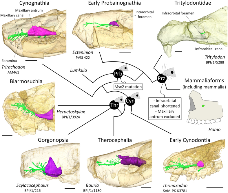
Figure 2. The evolution of the bony structures associated with the infraorbital nerve in Therapsida.
Digital reconstruction of the maxillary canal (in green) and maxillary antrum (in purple) based on CT scan images (see Material and Methods). Scale bars = 10 mm. Phylogeny after references Rubidge and Sidor31 and Liu and Olsen33. Lateral views of the skulls with bones transparent. Rostral is to the left. Videos of the CT images of the specimens are available in the Supplementary Information. Abbreviations : Cyn, Cynodontia clade; Prb, Probainognathia clade; Prz, Prozonstrodontia clade; Thr, Therapsida clade.

