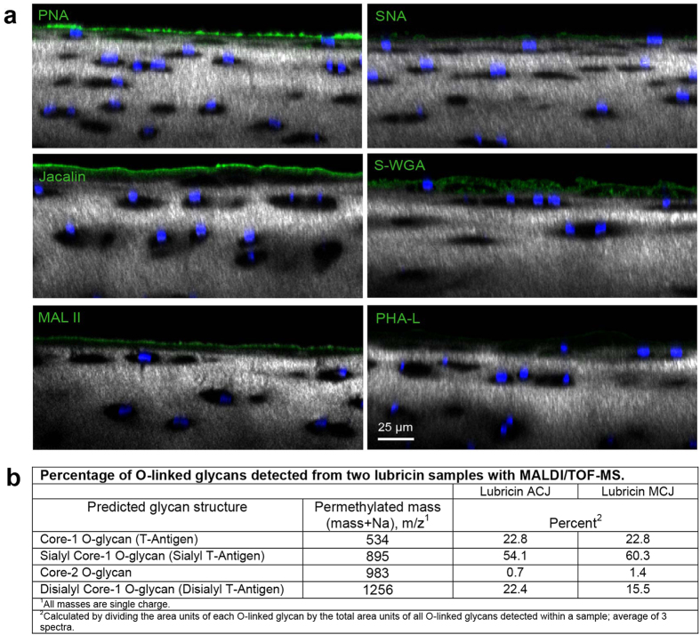
Figure 1. The glycophenotype of lubricin.
(a) Healthy equine articular cartilage imaged with confocal and multiphoton microscopy (40X) following incubation with fluorophore-conjugated lectins. Chondrocyte nuclei are stained with Hoechst 33342 (blue), and collagen is imaged using second harmonic generation microscopy (grey). PNA boundary layer staining indicates the presence of nonsialylated core-1 O-glycans (Galβ(1–3)GalNAc). Both MAL II, which preferentially binds to α2–3 sialylated core-1 O-glycans, and jacalin, which binds to both sialylated and nonsialylated core-1 O-glycans, labelled the boundary layer of articular cartilage. Faint staining of the superficial zone interterritorial matrix with S-WGA was present, whereas no appreciable boundary staining was present for either S-WGA or PHA-L, demonstrating that core-2 O-glycans and complex, branched N-glycans do not contribute substantially to the boundary layer oligosaccharide layer. Lectins: PNA, peanut agglutinin; jacalin; MAL II, Maackia amurensis lectin II; SNA, Sambucus nigra; S-WGA, S-wheat germ agglutinin; PHA-L,leucoagglutinin. (b) Relative ion intensity of O-linked oligosaccharides detected by MALDI/TOF-mass spectrometry in two healthy equine synovial fluid samples. Monosialylated structures predominate, followed by nearly equal distributions of disialylated and nonsialylated core-1 O-glycans.

