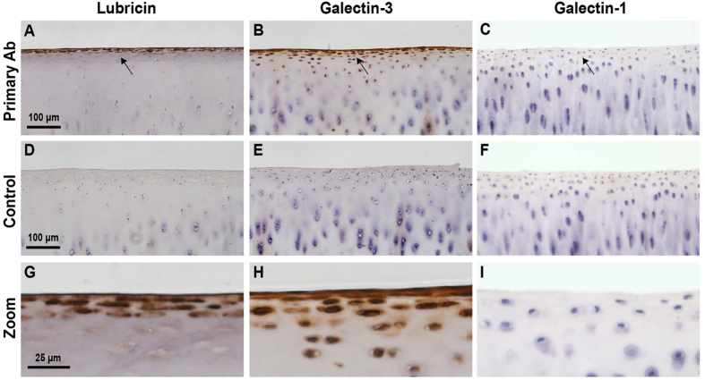
Figure 2. Lubricin and galectin-3 both localize to the cartilage surface.
Immunohistochemical detection of lubricin, galectin-3 and galectin-1 in photomicrographs of equine articular cartilage imaged at 20X (A–F) and magnified 4X (G–I). (A,G) Lubricin and (B,H) galectin-3 are both detected on the boundary layer of articular cartilage, whereas galectin-1 is not (C,I). Lubricin immunoreaction is observed in superficial zone chondrocytes and as a distinct layer along the lamina splendens (G), and galectin-3 immunoreaction is present within both superficial and middle zone chondrocytes and the lamina splendens (H). (D–F) Negative controls. Omission of primary antibody incubation confirms the absence of antigen-independent staining.

