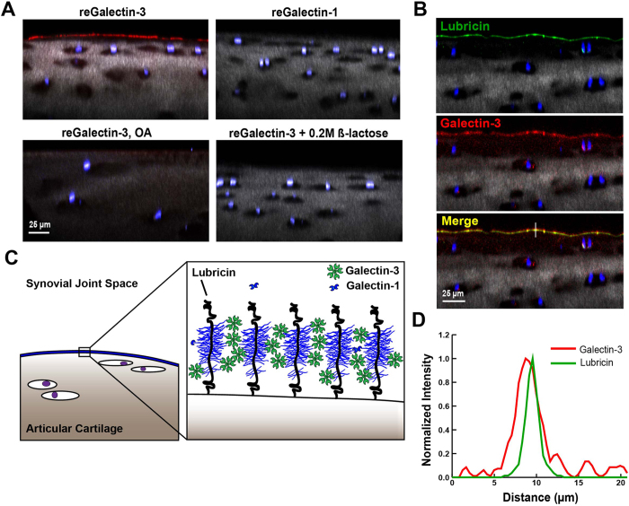
Figure 3. Galectin-3 binds to articular cartilage and colocalizes with lubricin.
(A) Equine articular cartilage explants imaged using confocal and multiphoton microscopy (40X) following incubation with Alexa647-conjugated galectins. Chondrocyte nuclei are stained with DAPI (blue), and collagen is imaged using second harmonic generation microscopy (grey). Galectin-3 prominently localizes to the boundary layer of healthy cartilage whereas galectin-1 does not. Galectin-3 staining is significantly decreased in joints with severe osteoarthritis (OA) and in the presence of 0.2 M β-lactose, suggesting carbohydrate-specific binding. (B) Lubricin stained with anti-lubricin mAb MABT401 and A647-conjugated galectin-3 binding to the surface of articular cartilage, demonstrating colocalization of lubricin and galectin-3. (C) Proposed role of galectin-3 in stabilizing the articular cartilage lubricin boundary layer. Pentavalent galectin-3 binds to glycans on adjacent lubricin polymer brushes, providing mechanical stabilization to the boundary layer through lubricin crosslinking. (D) Line scan from the white line in B demonstrating colocalization of lubricin and galectin-3 at the level of the articular cartilage boundary layer.

