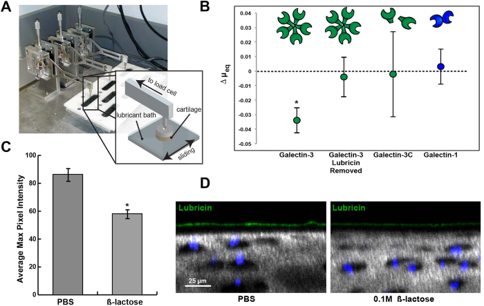
Figure 5. Galectin-3 enhances cartilage boundary lubrication.
(A) Schematic of custom tribometer used to measure boundary mode frictional coefficients for cartilage on glass in the presence of galectin solutions. (B) Galectin-3 decreases equilibrium friction coefficients as compared to paired controls (PBS), but only in the presence of endogenous articular lubricin. When lubricin is extracted using a 30-minute incubation with 1.5 M NaCl, galectin-3 no longer enhances lubrication. The galectin-3C mutant fails to enhance lubrication, suggesting that multimerization is critical for the ability of galectin-3 to facilitate boundary lubrication. Results are presented as mean ± standard deviation (SD) of n = 4. *p < 0.05. (C) Average maximum pixel intensity of boundary layer lubricin staining for cartilage explants incubated in either PBS or 0.2 M β-lactose for 12 hrs at 4 °C. Values represent the mean ± standard error (SE) of five independent samples quantified in NIH ImageJ software. Lubricin staining is decreased in the 0.1 M β-lactose treated explants, suggesting a role for galectins in stabilization of the lubricin boundary layer. (D) Equine articular cartilage explants incubated in either PBS or 0.2 M β-lactose for 12 hrs at 4 °C, followed by equilibration and incubation with α-lubricin mAb 9G3. Explants are imaged using confocal and multiphoton microscopy (40X). Chondrocyte nuclei are stained with DAPI (blue), and collagen is imaged using second harmonic generation microscopy (grey).

