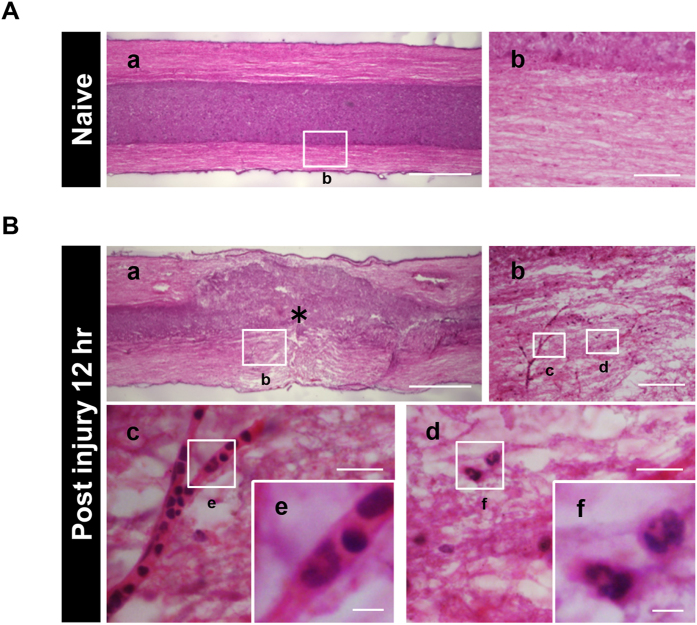
Figure 2. The structural integrity of the spinal cord is disrupted after SCI.
(A) Hematoxylin and eosin stained sagittal sections of the naive spinal cord showed the integrity of the neural structure, as well as a distinct demarcation between the white matter and gray matter. The image in (A–b) is a magnification of the boxed area in (A–a). (B) The neuronal structure was disrupted at 12 hours after SCI as shown by hematoxylin and eosin staining. The images in (B–b), (B–c), (B–d), (B–e) and (B–f) are magnifications of the boxed areas in (B–a), (B–b), (B–b), (B–c) and (B–d), respectively. The asterisk indicates the epicenter of the lesion. The images in (B–c) and (B–d) showed the blood vessel lumen and the characteristic lobulated nuclei of inflammatory cells. Scale bars (A–a,B–a): 500 μm; (A–b,B–b): 200 μm; (B–c,B–d): 50 μm; (B–e,B–f): 10 μm.

