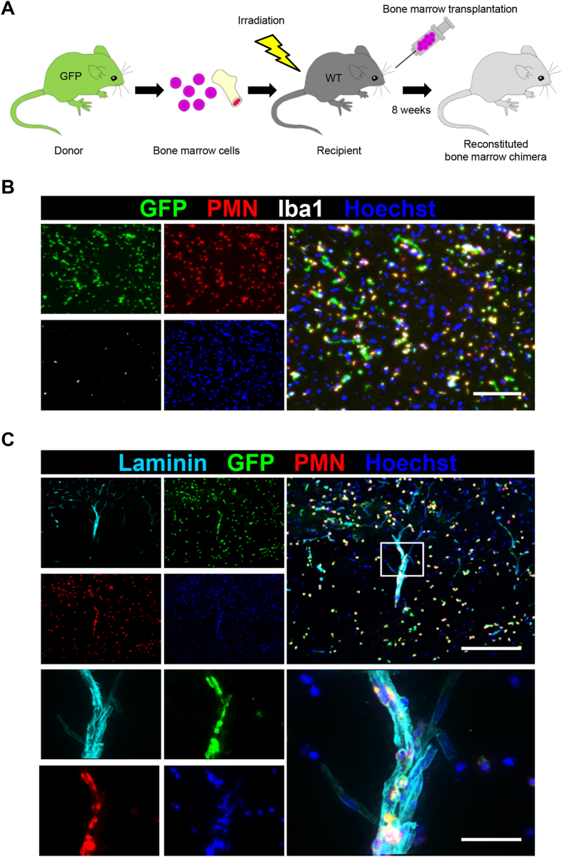
Figure 3. Peripheral blood-derived neutrophils infiltrate the injured spinal cord after SCI.
(A) A schematic illustration of the generation of bone marrow chimeric mice. Whole bone marrow was harvested from a green fluorescence protein (GFP) transgenic mouse donor. Bone marrow cell transplantation was performed, and the reconstituted bone marrow chimeric mice were analyzed at eight weeks after transplantation. (B) The immunohistochemical analysis of the injured spinal cord at 12 hours after SCI with GFP (green), PMN (red), Iba1 (white) and Hoechst (blue) staining. Almost all of the infiltrating peripheral blood-derived cells were PMN-positive neutrophils. (C) An immunohistochemical analysis of the injured spinal cord at 12 hours after SCI with laminin (light blue), GFP (green), PMN (red) and Hoechst (blue) staining. The lower images are magnifications of the boxed areas in the upper images. Neutrophils were observed inside the laminin-positive blood vessel walls. Scale bars (B): 100 μm; (C): 200 μm; inset: 50 μm.

