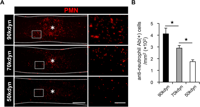
Figure 5. The number of infiltrating neutrophils in the injured spinal cord correlates with the severity of SCI.
(A) An immunohistochemical analysis of the injured spinal cord at 12 hours after SCI with PMN (red) staining in the mild (50 kdyn), moderate (70 kdyn) and severe (90 kdyn) SCI groups. The asterisk indicates the epicenter of the lesion. The right images are magnifications of the boxed areas in the left images. (B) The comparison of the number of infiltrating neutrophils in the injured spinal cord at 12 hours after SCI in the mild (50 kdyn), moderate (70 kdyn) and severe (90 kdyn) SCI groups, as determined by histological quantification. *P < 0.05, ANOVA with the Tukey-Kramer post-hoc test (n = 7 mice per group). Scale bars (A): 500 μm; insets: 100 μm.

