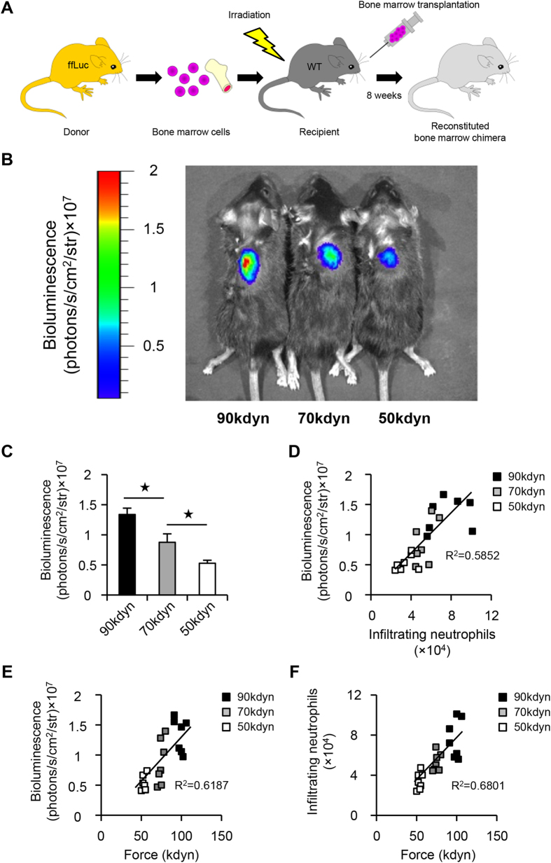
Figure 6. In vivo bioluminescence imaging predicts the functional prognosis after SCI.
(A) A schematic illustration of the generation of bone marrow chimeric mice. Whole bone marrow was harvested from a firefly luciferase (ffLuc) transgenic mouse donor. Bone marrow cell transplantation was performed, and the reconstituted bone marrow chimeric mice were analyzed at eight weeks after transplantation. (B) Representative pictures of bioluminescence imaging of the injured spinal cord at 12 hours after SCI in the mild (50 kdyn), moderate (70 kdyn) and severe (90 kdyn) SCI groups. (C) The bioluminescence signal intensity in the injured spinal cord correlated with the severity of SCI. *P < 0.05, ANOVA with the Tukey-Kramer post-hoc test (n = 7 mice per group). (D) The correlation between the number of infiltrating neutrophils as measured by flow cytometry and the bioluminescence signal intensity as measured by in vivo imaging (n = 7 mice per group, P < 0.0001, Spearman’s rank correlation coefficient). (E) The correlation between the severity of SCI and the bioluminescence signal intensity (n = 7 mice per group, P < 0.0001, Spearman’s rank correlation coefficient). (F) The correlation between the severity of SCI and the number of infiltrating neutrophils (n = 7 mice per group, P < 0.0001, Spearman’s rank correlation coefficient).

