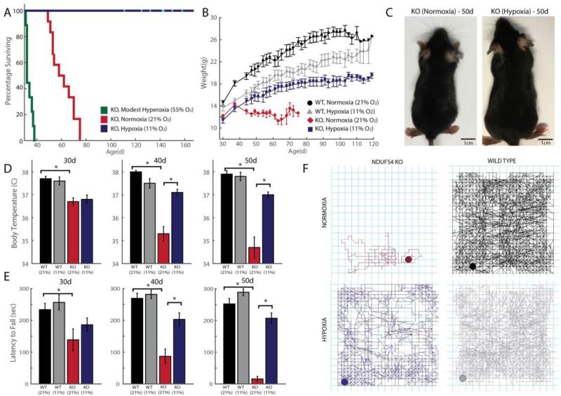
Figure 5. Chronic hypoxia extends lifespan and alleviates disease in a mouse model of Leigh syndrome, whereas chronic hyperoxia exacerbates disease.
(A) Ndufs4 KO mice of both genders were chronically exposed to hypoxia (11% O2), normoxia (21% O2) or hyperoxia (55% O2), at 30d of age and survival was recorded (n = 12, n = 12, n = 9 mice respectively). Cyan bars represent current age of hypoxic KO mice. (B) Body weights were measured in WT and KO mice exposed to normoxia or hypoxia, three times a week upon enrollment in the study. Weights are shown as mean ± S.E. (C) Representative images of 50d-old KO mice exposed to normoxia or hypoxia. (D) Body temperature was measured in KO mice exposed to normoxia or hypoxia at age ~30d, 40d and 50d. Temperatures are shown as mean ± S.E. (n ≥ 7 for all groups) (E) Latency to fall on an accelerating rod was measured as median values of triplicate trials per mouse for WT and KO mice, exposed to normoxia or hypoxia at different ages (n ≥ 7 for all groups). (F) Representative 1h locomotor activity traces of sick, normoxia-treated KO mice and age-matched hypoxia-treated KO mice, as well as controls. All data shown as normoxia KO (maroon), hypoxia KO (blue), normoxia WT (black) and hypoxia WT (grey). *denotes t-test p-value < 0.05.

