
Fig. 13.
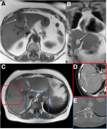
Axial black blood imaging (a) and coronal bright blood imaging (b) showing a well-defined low and high signal entity respectively consistent with a simple liver cyst. Axial black blood imaging (c) showing ill-defined low signal entity in the right lobe of liver (red box) which demonstrated features consistent with a metastasis on contrast enhanced CT (d). There is also a low signal entity in the vertebral body (blue box) which was confirmed to represent a sclerotic metastasis on CT (e)