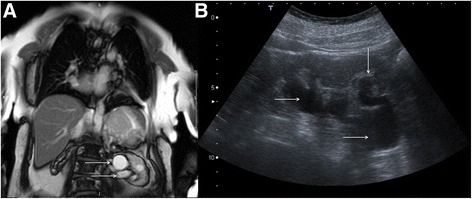
Fig. 16.

Coronal bright blood imaging (a) demonstrating a cystic entity at the left renal pelvis (solid white arrows). There is also a simple cortical renal cyst in the left lower pole. Correlative trans-abdominal ultrasound image (b) revealed the hypoechoic areas at the renal hilum are in continuity with a dilated proximal ureter and there is calyceal blunting (solid white arrows). These findings are consistent with hydronephrosis rather than para-pelvic cysts
