
Fig. 17.
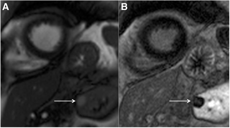
Short-axis steady state free precession cine image (a) revealing a high signal cyst in the visualized left kidney (solid white arrow). On the corresponding magnitude late gadolinium enhancement image (b) there is no enhancement of the cyst (solid white arrow) but diffuse enhancement of the surrounding normal renal parenchyma. This is a reassuring feature of renal cystic lesions