
Fig. 19.
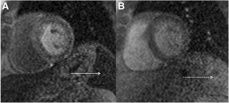
Real-time first pass myocardial perfusion at peak stress following administration of intravenous adenosine (a) revealing no discernible enhancement of the visualized spleen (solid white arrow). Real-time first pass myocardial perfusion at rest in the same patient (b) revealing normal diffuse enhancement of the splenic tissue in the field of view (dashed white arrow)