
Fig. 6.
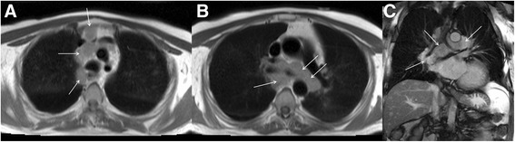
Axial black blood imaging (a and b) and coronal bright blood imaging (c) demonstrating florid mediastinal adenopathy engulfing the trachea and proximal main bronchi (solid white arrows) detected incidentally at the time of CMR
Official websites use .gov
A
.gov website belongs to an official
government organization in the United States.
Secure .gov websites use HTTPS
A lock (
) or https:// means you've safely
connected to the .gov website. Share sensitive
information only on official, secure websites.
Axial black blood imaging (a and b) and coronal bright blood imaging (c) demonstrating florid mediastinal adenopathy engulfing the trachea and proximal main bronchi (solid white arrows) detected incidentally at the time of CMR