
Fig. 9.
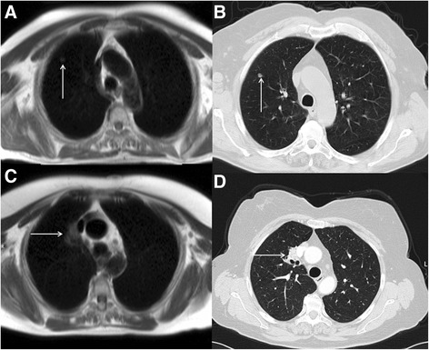
Axial black blood imaging (a) revealing an incidental 1.7 cm soft tissue density nodule in the right upper lobe (solid white arrows), which was subsequently confirmed with CT (b). Axial black blood imaging (c) demonstrating an incidental 3.1 cm soft tissue mass intimately related to the superior vena cava (solid white arrows), which was subsequently confirmed with CT (d)