Figure 1.
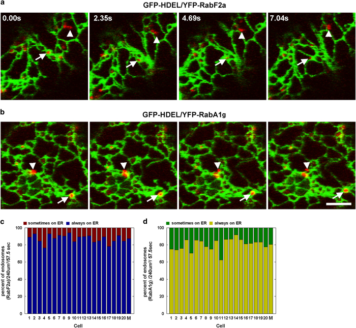
ER and endosomes are associated in plant cells. (a, b) Time-lapse microscopy in the cortical region of a Nicotiana tabacum leaf epidermal cells expressing GFP-HDEL (ER lumen) and either YFP-RabF2a (late endosome/MVBs) or YFP-RabA1g (post-TGN/EE late endosome/pre-vacuolar compartment) reveals that the majority of the endosomes are associated with the ER membranes over time (arrow) or only for a limited time (arrowhead). (c, d) Quantification of the ER–endosome association over time in individual cells (N=20) co-expressing GFP-HDEL and either YFP-RabF2a or YFP-RabA1g. The graph represents the percentage of endosomes that are continuously associated with the ER (always on ER) or are associated only for a limited number of frames (sometimes on ER) during the entire time-lapse sequence (57.5 s). The association was estimated for a total of 689 of RabF2a-labeled endosomes and 1165 RabA1g-labeled endosomes. The average of the percentages estimated for the 20 cells is also presented (M). Scale bars, 5 μm. ER, endoplasmic reticulum; MVBs, multivesicular bodies.

