Figure 2.
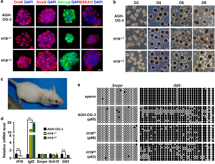
Characteristics of H19ΔAG-haESCs. (a) Immunofluorescence staining of AGH-OG-3 and H19ΔAG-haESCs. Scale bar, 20 μM. (b) Different days of embryoid bodies (EB) formation of AGH-OG-3 and H19ΔAG-haESCs. Scale bar, 200 μM. (c) Adult chimeric mouse produced by microinjection of haploid H19Δ1AG-haESC into diploid blastocysts. (d) Expression of imprinted genes measured by quantitative reverse transcription PCR (RT-qPCR). AGH-OG-3 AG-haESCs were used as control. Error bars, ±s.d. n=3. **P<0.01. (e) Methylation analysis of the Snrpn and Gtl2 DMRs in H19ΔAG-haESCs. Sperm DNA was used as control. Open circles represent unmethylated CpG sites, whereas filled circles represent methylated CpG sites.

