
Figure 6.
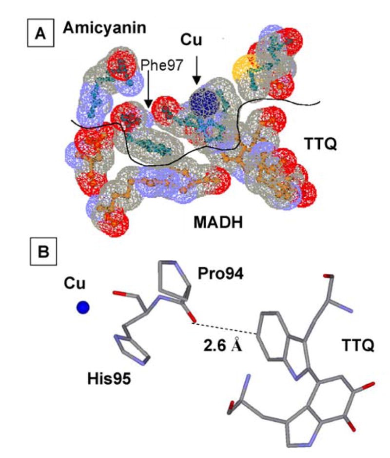
The amicyanin-MADH interface. A. Residues of amicyanin (green) and MADH (brown) near the site of interprotein ET are shown as ball and stick with their van der Waals radii colored as oxygen (red), nitrogen (blue), carbon (grey) and sulfur (yellow). B. Predicted point of interprotein ET from the backbone oxygen of Pro54 of amicyanin to TTQ.