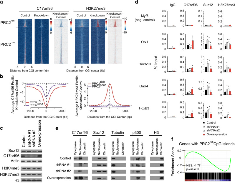
Figure 2.
C17orf96 depletion affects Suz12 chromatin binding and H3K27me3 deposition. (a) Heatmap at CGIs (as in Figure 1e) of the ChIP-seq of C17orf96 and H3K27me3 in mES cells infected with scrambled shRNA (‘control’) or a specific shRNA for C17orf96 (shRNA #2; ‘knockdown’). The last heatmap for each antibody shows the subtraction of the knockdown versus control cells. (b) Differential profiles of knockdown versus control. Knockdown of C17orf96 reduces C17orf96 signal at PRC2-poor and PRC2-rich CGIs, but increases the H3K27me3 specifically at PRC2-rich CGIs. (c) Western blot analysis of four created cell lines, infected with a construct expressing control shRNA, two specific shRNA for mouse C17orf96 and mouse C17orf96 (untagged; ‘overexpression’). No global change of Suz12, H3K4me3 and H3K27me3 could be detected. See also Supplementary Figure S2. (d) ChIP experiments on genes with PRC2-rich CGIs, using the four cell lines described. Knockdown of C17orf96 enhances Suz12 and H3K27me3 levels. Overexpression reduces Suz12 levels, but does not appear to affect H3K27me3. Values represent the average and s.d. of two independent experiments. *P<0.05. (e) Cellular fractionation of the four cell lines. The level of chromatin-bound Suz12 negatively correlates with the level of C17orf96. Notably, C17orf96 is mostly in the nucleoplasm and cytoplasm, but hardly in the chromatin fraction. See also Supplementary Figure S5D. (f) Gene set enrichment analysis of genes possessing a PRC2-rich CGI using microarray data from De Cegli et al. [16]. Knockdown of C17orf96 leads to a significant reduced expression of those genes.

