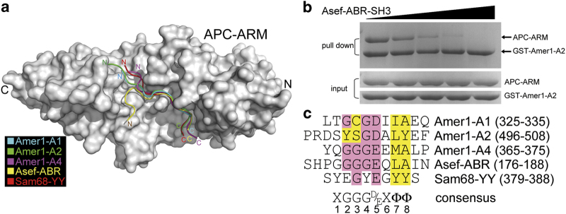
Figure 7.
Comparison of the binding patterns of Amer1-A1, -A2, -A4, Asef-ABR, and Sam68-YY reveals a common recognition motif for APC–ARM association. (a) Structural superimposition of APC–ARM in complexes with its binding partners (Amer1-A1: cyan, Amer1-A2: green, Amer1-A4: magenta, Asef-ABR: yellow, and Sam68-YY: red). (b) Competition between Asef-ABR-SH3 and Amer1-A2 for binding to APC–ARM using the GST pull-down assay. (c) Structure-based alignment of the APC-binding sequences of human Amer1-A1, -A2, -A4, Asef-ABR, and Sam68-YY. The consensus APC–ARM binding motif XGGGD/EXΦΦ (X stands for any residue, and Φ represents a hydrophobic residue) is shown below the sequences.

