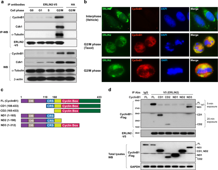
Figure 4.
Endoplasmic reticulum lipid raft-associated protein 2 (ERLIN2) complexes with Cyclin B1, Cdk1 and α-tubulin in G2/M phase. (a) IP–western blot (IP–WB) analysis of the interactions between ERLIN2, Cyclin B1, Cdk1 or α-tubulin in ERLIN2 (tagged with V5)-expressing CHO cells synchronized at G0, G1, S or G2/M phase. Total cell lysates were immunoprecipated with the anti-V5 antibody or anti-HA (negative control). The immunoprecipated proteins were subjected to immunoblotting analysis using the anti-Cyclin B1, anti-Cdk1 or anti-α-tubulin antibody. The levels of Cyclin B1, Cdk1, α-tubulin and β-actin in total cell lysates were determined as the controls. (b) Immunofluorescent (IF) staining of ERLIN2 (green fluorescence) and Cyclin B1 (red fluorescence) in the CHO cell line stably expressing ERLIN2. The cells were treated with taxol (10 μm) or the vehicle dimethyl sulfoxide (DMSO) for 1 h. Nuclei were stained with DAPI (blue). The images on the right were for a representative CHO cell in G2/M phase stained for ERLIN2, Cyclin B1 and nuclei. Magnification: ×400. (c) Domain structures of full-length (FL) Cyclin B1 protein and its truncated mutants. Cyclin B1 contains an N-terminal destruction box (DB), followed by a cytoplasmic retention sequence (CRS) and a cyclin box domain. The amino acid number of each isoform was indicated. (d) IP–western blot analysis of the interactions between ERLIN2, Cyclin B1 (FL), and Cyclin B1 truncated isoforms (illustrated in c) in CHO cells. Plasmid vectors expressing flag-tagged Cyclin B1 and truncated Cyclin B1 isoforms were transfected into the CHO cell line stably expressing V5-tagged ERLIN2. Total cell lysates from transfected CHO cells were immunoprecipated with the anti-V5 antibody or rabbit IgG (negative control). The pull-down proteins were probed with the anti-flag antibody to detect the association of ERLIN2 with Cyclin B1 or its truncated forms. The pull-down proteins were probed with the anti-V5 antibody for the loading controls. Because of the different strengths of interactions between ERLIN2 and the Cyclin B1 truncated forms, both short (5 min)- and long (20 min)-time film exposure images were included to identify the interaction signals. The levels of Cyclin B1 or its truncated forms and GAPDH in total cell lysates were determined as the controls (lower panels).

