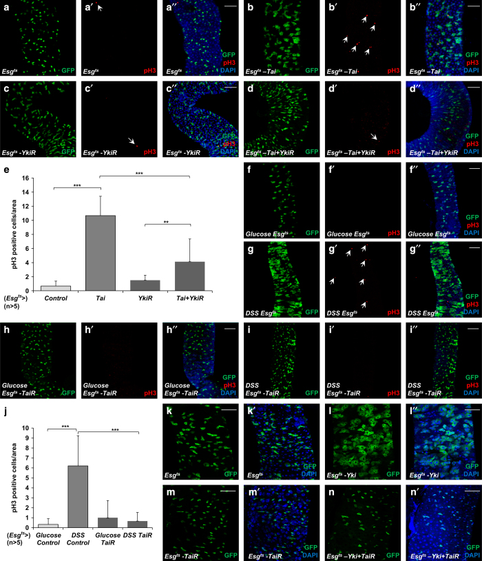
Figure 8.
Tai is required for ISC proliferation. (a–d′′) Midguts from adult flies of indicated genotypes were stained with pH3 antibody (red) and DAPI. esg-GFP marked the ISCs/enteroblasts (EBs) and pH3-positive cells were indicated with arrows. Scale bars, 50 μm. (e) The comparison of the number of pH3-positive cells shown in a–d′′. The data were quantified using an unpaired t-test. The results represented the mean+s.e.m. ***P<0.001, **P<0.01, *P<0.1, (n>5) for each genotype. (f–i′′) Midguts from adult flies of indicated genotypes treated with glucose or DSS were stained with pH3 antibody (red) and DAPI. esg-GFP marked the ISCs/EBs and pH3-positive cells were indicated with arrows. Scale bars, 50 μm. (j) The comparison of the number of pH3-positive cells shown in f–i′′. The data were quantified using an unpaired t-test. The results represented the mean+s.e.m. ***P<0.001, **P<0.01, *P<0.1, (n>5) for each genotype. Scale bars, 50 μm. (k–n′) Midguts from adult flies of indicated genotypes were stained with DAPI. esg-GFP marked the ISCs/EBs. Scale bars, 50 μm.

