
Figure 7.
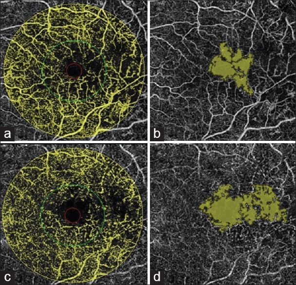
Image of a patient with mild non-proliferative diabetic retinopathy and diabetic macular edema. (a and b) Showing the en-face image of the superficial plexus with distortion of foveal avascular zone (FAZ). (c and d) Showing the en-face image of the deep plexus with distortion of FAZ, more profound than in superficial plexus. This happens due do accumulation of fluid in retinal layers.