
Figure 9.
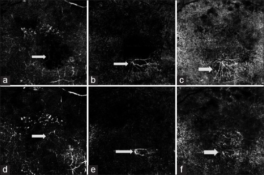
Optical coherence tomography angiography (OCTA) images of a patient with choroidal neovascularization (CNV) before (a.c) and 2 weeks after treatment with anti VEGF (d.f). (a) OCTA image of the deep plexus, with cystic changes causing obliteration of vascular reflection. (b) NV membrane in the photoreceptor zone. (c) NV membrane extending in the deeper choroid. (d) OCTA of the deep retinal plexus after treatment with anti-VEGF. Vessels in the deeper retinal plexus are better visualized due to regression of cystic changes. (e) Regression of the NV membrane in the photoreceptor zone. Obliteration of smaller vessels and decrease in the size of main feeder trunk is noted. (f) Regression in the size of NV membrane is also noted in the choriocapillary layer.