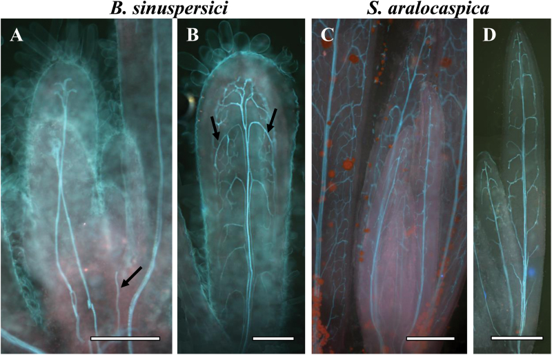
Fig. 1.
Cleared young leaves of Bienertia sinuspersici (A, B) and Suaeda aralocaspica (C, D) viewed under UV light at different stages of vein initiation. There is acropetal formation of the central vein towards the leaf tip (illustrated by arrow in panel A) and basipetal direction of lateral vascular vein development from the tip to base of the leaf (illustrated by arrows in panel B). Scale bars: 250 μm for A, B; 500 μm for C, D.

