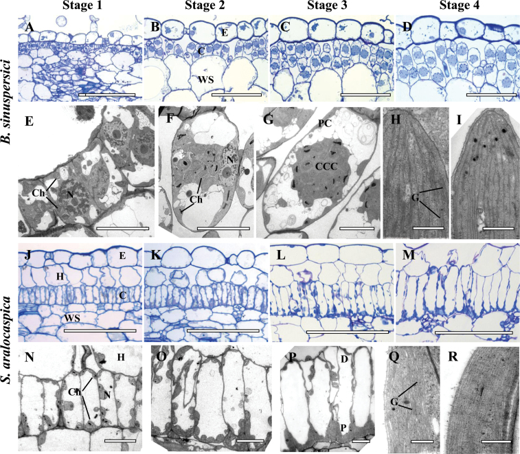
Fig. 2.
Light and electron microscopy of Bienertia sinuspersici (A–I) and Suaeda aralocaspica (J–R) with longitudinal sections of young leaves. The sections show a basipetal developmental gradient with gradual structural differentiation of SC-C4 chlorenchyma cells with four stages along the longitudinal gradient: Stage 1 (A, E, J, N), Stage 2 (B, F, K, O), Stage 3 (C, G, L, P) and Stage 4 (D, H, I, M, Q, R). Panels A–D from B. sinuspersici and J–M from S. aralocaspica are light microscopy micrographs of longitudinal sections showing the development of chlorenchyma cell lineages from the base (A, J) to the tip (D, M) of young leaves, with the direction of maturation from left to right. Panels E–G from B. sinuspersici and N–P from S. aralocaspica, are TEM micrographs showing internal structural development within a single chlorenchyma cell along the longitudinal gradient from the base (E, N) to the middle region (G, P) of a young leaf. Panels H, I, show the ultrastructure of chloroplasts in the central cytoplasmic compartment, CCC (panel H) and the periphery (panel I) within a single chlorenchyma cell at the tip of a young B. sinuspersici leaf. Panels Q, R show ultrastructure of chloroplasts within the proximal (Q) and distal (R) regions of chlorenchyma cell at the tip of a young S. aralocaspica leaf. E, epidermis; C, chlorenchyma; CCC, central cytoplasmic compartment; D, distal end; G, grana; H, hypoderm; N, nucleus; P, proximal end; WS, water storage. Scale bars: 100 μm for A–D and J–M; 10 μm for E–G and N–P; 0.5μm for H, I, Q, R. (This figure is available in color at JXB online.)

