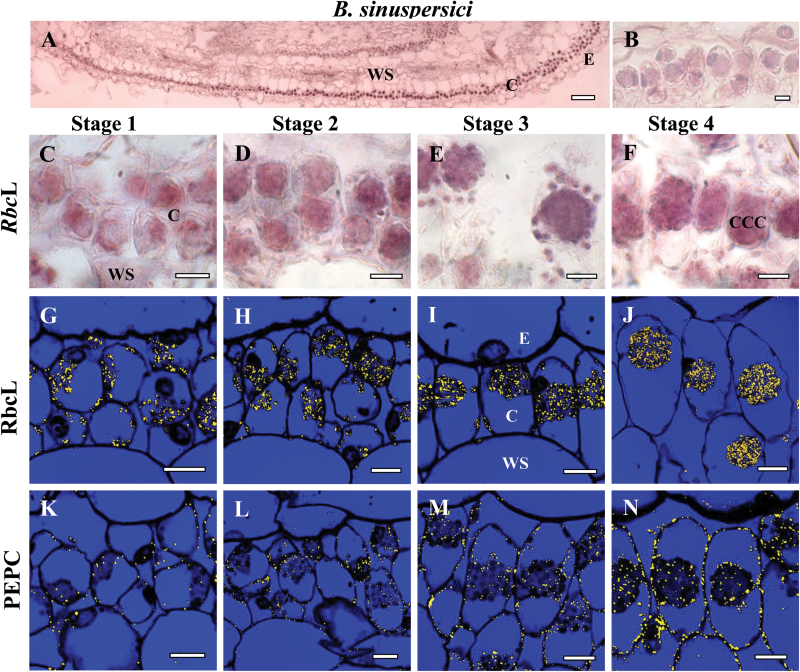
Fig. 3.
In situ hybridization of rbcL mRNA (A–F) and in situ immunolocalization of Rubisco rbcL (G–J) and PEPC (K–N) with longitudinal sections of young leaves from base to tip of Bienertia sinuspersici at four stages of development: Stage 1 (C, G, K), Stage 2 (D, H, L), Stage 3 (E, I, M) and Stage 4 (F, J, N). The dark purple signal indicates the specific hybridization to an antisense mRNA probe for rbcL mRNA (panels A, C–F). Yellow particles (panels G–N) indicate labeling with rbcL and PEPC antibodies. (A) Basipetal gradient of rbcL transcript accumulation from base (left) to tip (right). (B) Sense probe control showing the very low background staining that occurred in mRNA sense strand hybridization reactions. E, epidermis; CCC, central cytoplasmic compartment; C, chlorenchyma; WS, water storage. Scale bars: 100 μm for A; 10 μm for B–N.

