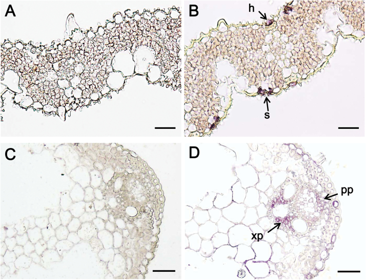
Fig. 5.
PEPCK is present in the stomata, s, hydathode, h, and vascular tissue (xylem parenchyma, xp; phloem parenchyma, pp). Transverse sections of rice after immunodiazostaining with pre-immune (A and C) and PEPCK serum (B and D). Serum used in panel B was a peptide antibody to the chromosome 3 isoform in rice. Serum used in panel D was a protein antibody to PEPCK in cucumber that was more sensitive to PEPCK located in the vascular tissue but less sensitive to PEPCK located in the stomata and hydathode of rice. Bars: 20 μm for A, B; 30 μm for C, D.

