
Figure 4. Thalamic excitation of SST interneurons is reduced during development.
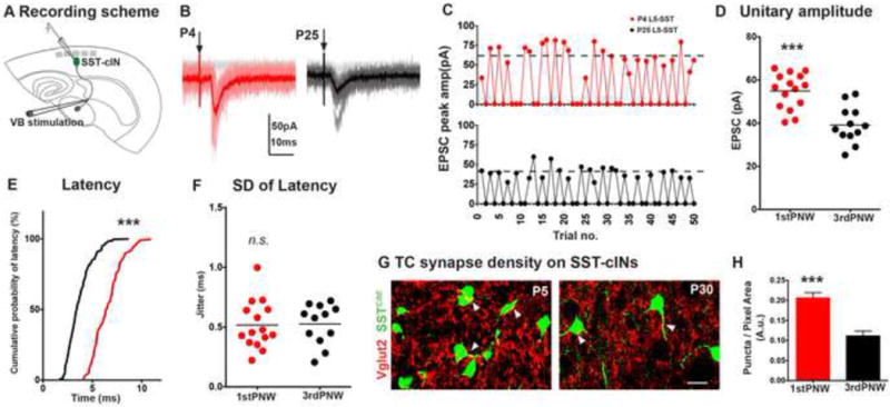
(A) Synaptic events in SST interneurons recorded from L5B/L6A in a TC slice preparation at P3–6 and P20–25 using minimal stimulation protocol (50–500μA) to evoke EPSCs only in half of the trials. (B) Representative traces of TC-EPSCs in P4 (red) and P25 (black) SST interneurons. (C) Exemplary recording sessions showing distribution of minimal events during 50 sweeps, dashed lines represent the averaged potency. (D) Excluding failures, peak TC EPSCs in SST interneurons of immature brains (n=15) showed a small but significant improvement over mature SST interneurons than those of older mice (n=12, p<0.0001, Mann-Whitney test) (E). Thalamic EPSC latency was longer in immature mice, relative to older animals (p<0.0001, Kolmogorov-Smirnov test). (F) Mean standard deviation of latency was not significantly different between two ages (p>0.05, Mann-Whitney test). For each time point, n ≥ 4 animals, n ≥ 2 brain slices were analyzed per animal. See also Figure S4. (G) Vglut2 immunoflouroscence in genetically identified P5 (left), and P30 SST interneurons (right). Images are pseudocolored for clarity, scale bar corresponds to 10μm. (H) Vglut2 synaptic puncta density on SST interneurons were significantly larger in P5 animals compared to older animals (p<0.0001). n ≥ 9 areas analyzed from n ≥ 10 cells, 3 animals, 3 sections per animal, mean ± SEM, Mann-Whitney test.