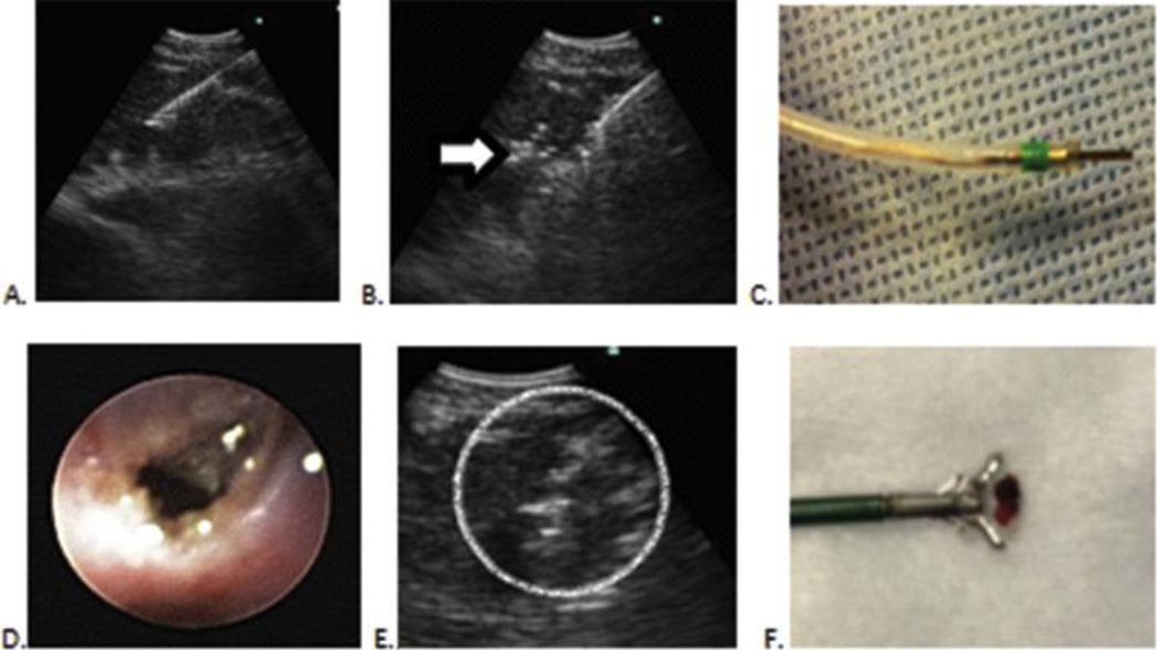
Figure 1. Technique employing EBUS-guided cautery-assisted forceps biopsies of lymph nodes.
A. EBUS-TBNA needle in lymph node
B. EBUS view with cautery knife inside same lymph node. Note mild artifact (Arrow)
C. Electrocautery knife that is inserted through working channel of EBUS scope
D. Mucosal incision seen after electrocautery incision.
E. Forceps seen inside lymph node (Circle)
F. Specimen obtained by forceps for surgical pathology

