
Figure 2. mosGCTLs facilitate the colonization of the A. aegypti midgut by gut bacteria.
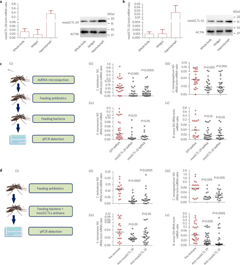
a,b, Distribution of mosGCTL-29 (a) and mosGCTL-32 (b) in A. aegypti. The abundance of mosGCTLs was detected in the whole bodies, midguts and haemolymph via qPCR (left panels) and western blotting (right panels). For the western blotting experiment, the mosquito whole bodies, midguts and haemolymph were dissected and lysed for detection. The polyclonal antibodies against mosGCTL-29 and mosGCTL-32 were generated in mice. The detection of actin served as an internal control. A total of 100 μg of protein lysate was loaded in each lane. Data in a,b are represented as mean±s.d. in each group. c, Knockdown of mosGCTLs impaired colonization of gut bacteria in the A. aegypti midgut. (i) Schematic representation of the study design. mosGCTL-29 and mosGCTL-32 were silenced in antibiotic-treated mosquitoes. The GFP dsRNA-treated mosquitoes served as mock controls. Subsequently, 1 optical density (OD) of C. testosteroni (ii), C. meningosepticum (iii), S. marcescens (iv) and B. cereus (v) was fed to the mosquitoes 3 days after dsRNA inoculation, respectively. d, Immuno-blockade of mosGCTLs reduced colonization of gut bacteria in the A. aegypti midgut. (i) Schematic representation of the study design. The antibodies against mosGCTL-29 or mosGCTL-32 were fed together with 1 OD of C. testosteroni (ii), C. meningosepticum (iii), S. marcescens (iv) and B. cereus (v) to the antibiotic-treated mosquitoes. Mosquitoes fed a pre-immune antibody served as the mock control. c,d, The bacterial burden was determined by SYBR Green qPCR. The qPCR primers for bacterial 16S rDNA are described in Supplementary Table 5. One dot represents one mosquito gut. The horizontal line represents the median value of the results. Data were analysed using the non-parametric Mann–Whitney test. The results were combined from at least two biologically independent experiments.