
Figure 4. mosGCTLs and AMPs are simultaneously regulated by the Imd pathway.
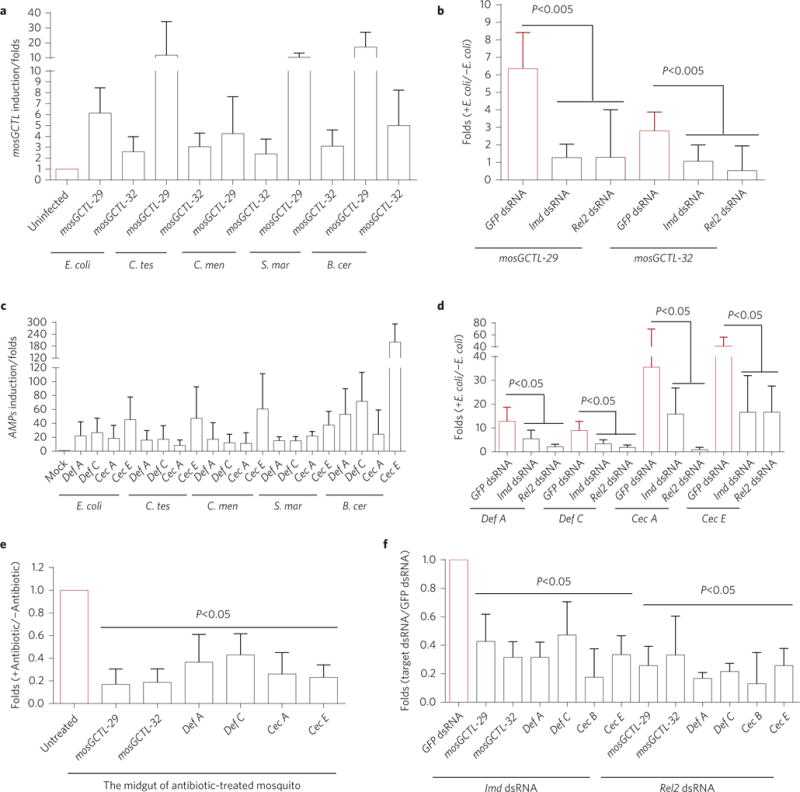
a,c, The expression of mosGCTLs and AMPs were simultaneously induced by systemic inoculation with E. coli, C. testosteroni (C. tes), C. meningosepticum (C. men), S. marcescens (S. mar) and B. cereus (B. cer). The mock-treated mosquitoes received inoculations of PBS buffer. The mRNA abundance of mosGCTLs (a) and AMPs (c) was assessed 4 h after bacterial inoculation. The induction is presented as the fold-change relative to that in the mock mosquitoes without bacteria treatment. b,d, The role of the Imd signaling pathway in mosGCTL and AMP induction. The key factors in the Imd pathway (Imd and Rel2) were silenced in A. aegypti. A 0.005 OD aliquot of E. coli in PBS was sequentially inoculated 3 days later, and the expression of inducible mosGCTLs (b) and AMPs (d) was assessed 4 h after bacterial inoculation. Mosquitoes inoculated with GFP dsRNA and subsequently infected with E. coli were included as controls. The induction is presented as the fold-change relative to that in the uninfected mosquitoes. e, Removal of gut commensal bacteria suppressed the expression of mosGCTLs and AMPs in the midguts. The expression of mosGCTLs and AMPs was determined in the midguts of antibiotic-treated mosquitoes. Mosquitoes without antibiotic treatment served as mock controls. f, Knockdown of the Imd and Rel2 genes reduced the expression of mosGCTLs and AMPs in the midguts. The Imd and Rel2 genes were silenced by dsRNA-mediated thoracic microinjection. Mosquitoes inoculated with GFP dsRNA were included as controls. After 3 days, the midguts of the treated mosquitoes were isolated to determine the mRNA abundance of mosGCTLs and AMPs. The result was read through qPCR and normalized to A. aegypti actin (AAEL011197). The qPCR primers are described in Supplementary Table 5. Data are represented as mean±s.d. in each group and analysed using the nonparametric Mann–Whitney test. The experiment was biologically repeated three times with similar results.