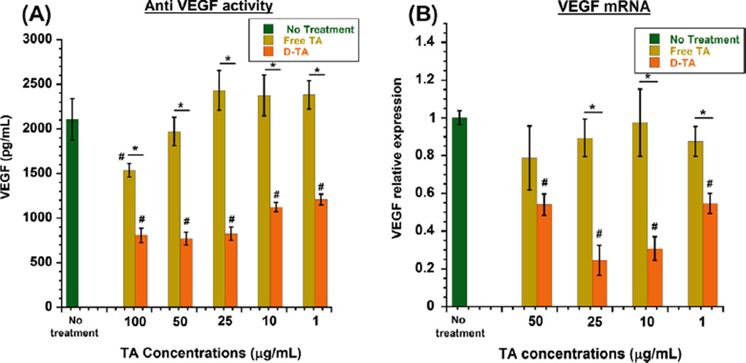
Fig. 5. Anti-VEGF activity of D-TA in human retinal pigment epithelial cells.
ARPE-19 (passage 21) were subjected to hypoxia for 6 h and treated with D-TA and free TA (equivalent drug basis) at indicated concentrations for 24 h and replaced with culture medium. 24 h post treatment VEGF was analyzed using human VEGF ELISA kit and RT-PCR for mRNA expression (A) VEGF secretion analysis in culture medium using ELISA (n = 6) and (B) VEGF mRNA expression levels relative to GAPDH using RT-PCR (n = 3) data denote mean ± SEM, *P < 0.01 vs free TA and #P < 0.01 vs group with no treatment. (For interpretation of the references to color in this figure legend, the reader is referred to the web version of this article.)

