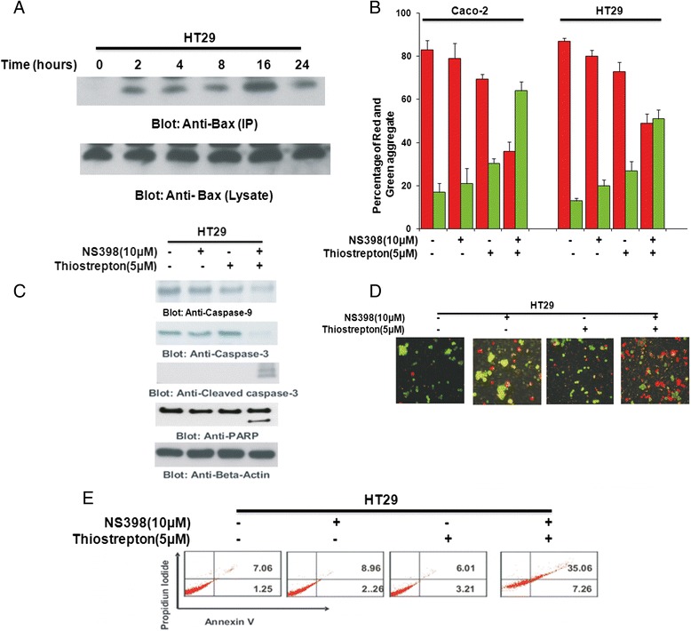
Fig. 4.

Combination of NS398 and Thiostrepton at sub-optimal doses induces caspase-dependent apoptosis via the mitochondrial pathway in CRC cells. a HT29 cells were treated with combination of 10 μM NS398 and 5 μM Thiostrepton for indicated time periods. Cells were lysed in 1 % Chaps lysis buffer and subjected to immuno-precipitation with anti-Bax 6A7 monoclonal antibody and probed with specific polyclonal anti-Bax antibody for detection of conformationally changed Bax protein. In addition, the total cell lysates were applied directly to SDS–PAGE, transferred to immobilon membrane and immuno-blotted with specific anti-Bax polyclonal antibody. b Caco-2 and HT29 cells were treated with 10 μM NS398 and 5 μM Thiostrepton either alone or in combination for 48 h. Live cells with intact mitochondrial membrane potential (red bars) and dead cells with lost mitochondrial membrane potential (green bars) was measured by JC-1 staining and analyzed by flow cytometry as described in Materials and Methods. The graph displays the mean +/- SD (standard deviation) of three independent experiments. c HT29 cells were treated with 10 μM NS398 and 5 μM Thiostrepton alone or in combination for 48 h. Cells were lysed and equal amounts of proteins were immunoblotted with antibodies against caspase-9, caspase-3, cleaved caspase-3, PARP, and Beta-actin. d Caco2 and HT29 cells were treated with 10 μM NS398 and 5 μM Thiostrepton either alone or in combination for 48 h and cell death was analyzed by Live/Dead Assay. e HT29 cells were treated with 10 μM NS398 and 5 μM Thiostrepton alone or in combination for 48 h. Thereafter, the cells were washed, and stained with annexin V/propidium iodide, and analyzed by flow cytometry as described in Materials and methods
