Fig. 1.

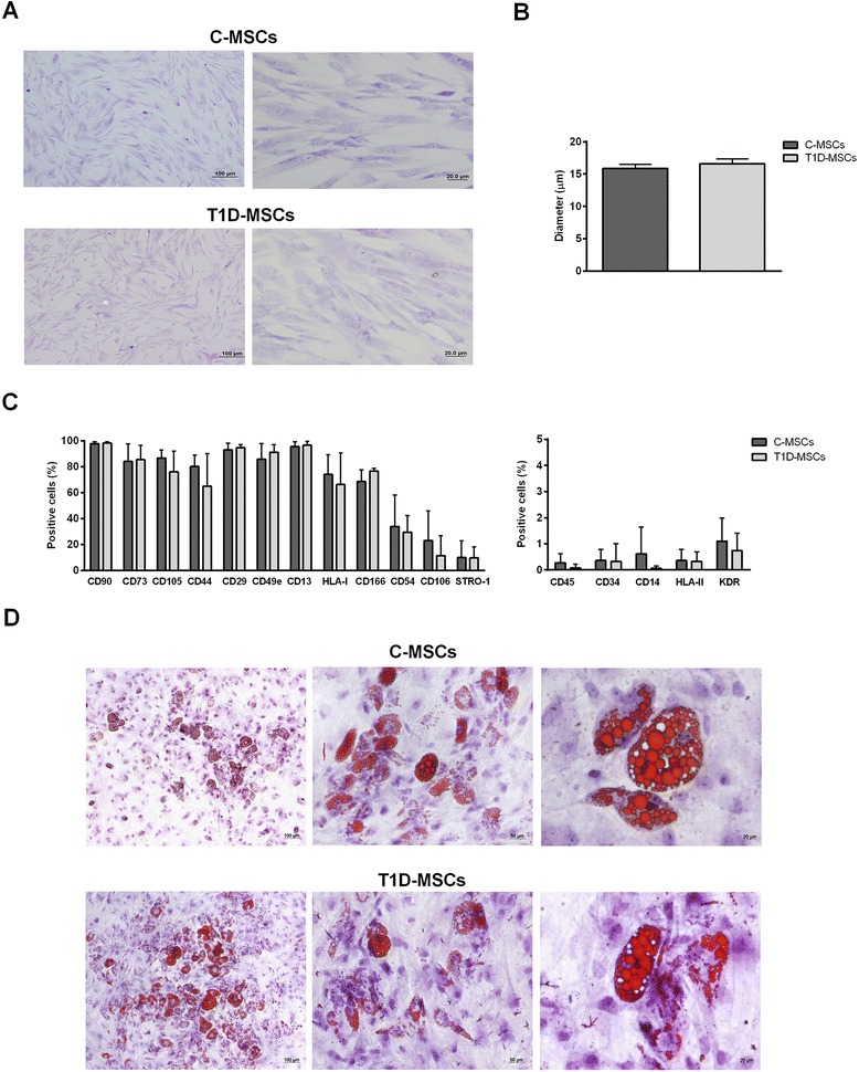
Characterization of T1D-MSCs isolated. a Morphological characterization. At the third passage, C-MSCs (upper panel) and T1D-MSCs (lower panel) showed homogeneous spindle-shaped fibroblast-like growth (Leishman staining, 100× and 400× magnification, respectively). b The diameter of C-MSCs (n = 5) and T1D-MSCs (n = 5) in suspension was determined by ViCell XR equipment (2000 cells/sample). c Expression of surface immunophenotypic markers. Graphs display the phenotype of MSCs in culture at the third passage. MSCs from T1D patients were phenotypically similar to those from healthy donors. Bars represent mean ± SD. d In vitro adipocyte differentiation. C-MSCs (upper panel) and T1D-MSCs (lower panel) were able to differentiate towards adipogenic lineage. The presence of lipid vacuoles in the cytoplasm was identified by Sudan II-Scarlet staining (100×, 200×, and 400× magnification, respectively). C-MSCs mesenchymal stromal cells from bone marrow of healthy individuals, T1D-MSCs mesenchymal stromal cells from bone marrow of newly diagnosed T1D patients
