Fig. 5.

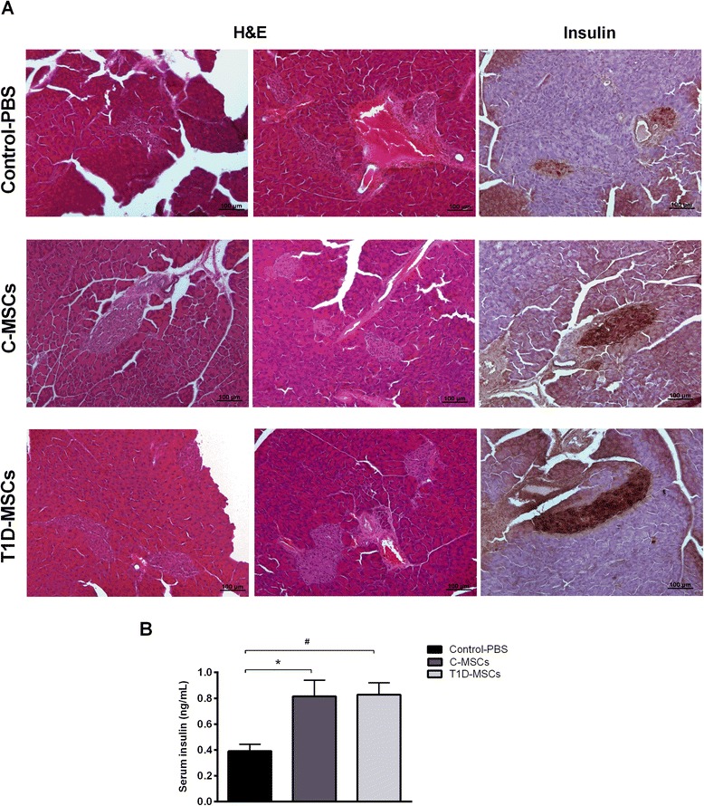
T1D-MSC transplantation reduces insulitis and augments insulin production by pancreatic β cells in diabetic-treated mice. a Pancreata from Control-PBS-treated, C-MSC-treated, and T1D-MSC-treated mice were collected 35 days after the treatments. Islet morphology was evaluated by H & E staining and the in situ insulin content was detected by immunohistochemistry analysis. Representative H & E or insulin-stained islets from the Control-PBS group (upper panel), C-MSC-treated group (middle panel), and T1D-MSC-treated group (lower panel) are shown. Original magnification: 100×. b Blood samples were collected on day 35 and circulating-insulin levels were determined by ELISA. Bars represent mean ± SD. *P <0.05 (Control-PBS × C-MSCs); # P <0.05 (Control-PBS × T1D-MSCs). C-MSCs mesenchymal stromal cells from bone marrow of healthy individuals, H & E hematoxylin and eosin, PBS phosphate-buffered saline, T1D-MSCs mesenchymal stromal cells from bone marrow of newly diagnosed T1D patients
