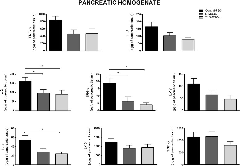
Fig. 7.

Intrasplenic T1D-MSC administration modulates proinflammatory cytokines in the pancreatic tissue of STZ-induced diabetic mice. Pancreata were obtained from Control and MSC-treated groups 35 days after treatment. The samples were weighed and homogenized in the presence of proteases inhibitor. Levels of IL-2, IL-4, IL-17, IL-6, IFNγ, TNFα, and IL-10 were measured by cytokine beads array (CBA) method. The TGF-β level was quantified by ELISA. Cytokine concentrations are represented by picograms of protein per gram of pancreatic tissue. Bars represent mean ± SD. *P <0.05 (Control-PBS × C-MSCs); # P <0.05 (Control-PBS × T1D-MSCs). C-MSCs mesenchymal stromal cells from bone marrow of healthy individuals, IFN interferon, IL interleukin, PBS phosphate-buffered saline, T1D-MSCs mesenchymal stromal cells from bone marrow of newly diagnosed T1D patients, TGF-β transforming growth factor beta, TNFα tumor necrosis factor alpha
