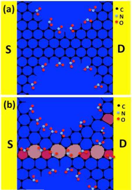
Figure 6. Crystal structures of gCH4 and gEtOH nanomeshes.
A structural representation (not to scale) comparing the defect densities between a, gCH4 and b, gEtOH nanomesh sensor devices, and adsorptions of NO2 molecules on the defect sites. A typical intrinsic defect (grain boundary) of polycrystalline gEtOH nanomesh labeled as colored unsaturated carbon (pentagons and heptagons) structure in b.

