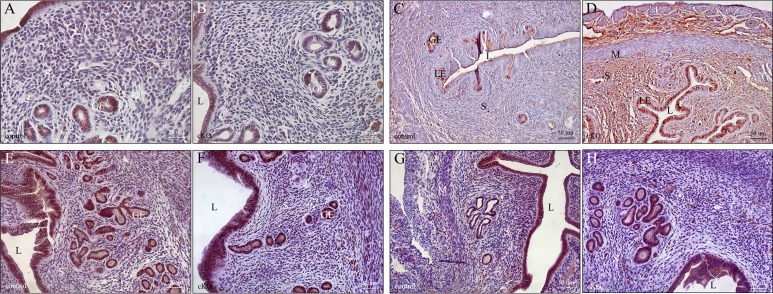
FIG. 5.
After 6-mo fertility studies, parous uteri from Arid1a cKO mice showed increased proliferation. ARID1A expression was present in >95% of epithelial cells from control (A) but only 50%–80% of epithelial cells from cKO (B) uteri. However, increased proliferation in both epithelium and stromal compartments was apparent in cKO (D) compared to control (C) parous uteri by Ki67 staining. Expression of ESR1was similar between control (E) and cKO (F) parous uteri. Similarly, expression of PGR was similar between control (G) and cKO parous uteri (H). GE, glandular epithelium; L, lumen; LE, luminal epithelium; M, myometrium; S, uterine stroma. Bars = 25 μm (A, B) and 50 μm (C–H).

