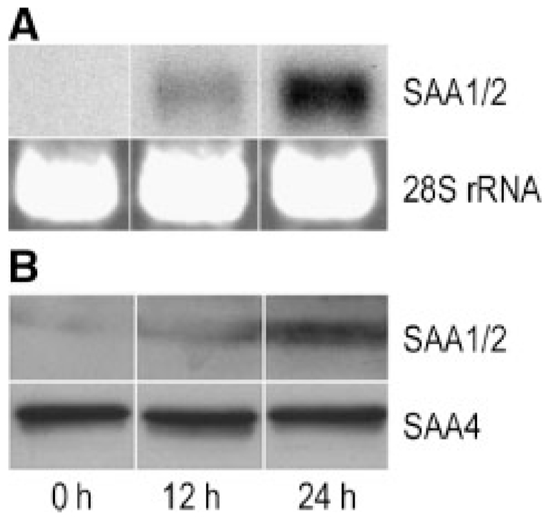
Fig. 4.
Time-dependent expression of SAA in osteosarcoma cells: MG-63 cells were stimulated with 10 ng/ml IL-1β for 12 and 24 h. A: RNA was isolated and Northern blot experiments were performed using SAA1/2 cDNA as a probe. The 28S rRNA was used as gel loading control. B: Cells were lysed and total cellular protein was subjected to SDS–PAGE and transferred to membranes. Western blot experiments were performed using sequence-specific anti-human SAA1/2 or SAA4 peptide antisera (see Materials and Methods section) as primary antibodies. One representative experiment out of three is shown.

